Sonography
Sonography, or ultrasound, is a non-invasive method for monitoring fetal development. We provide high-quality ultrasound services.
Book Appointment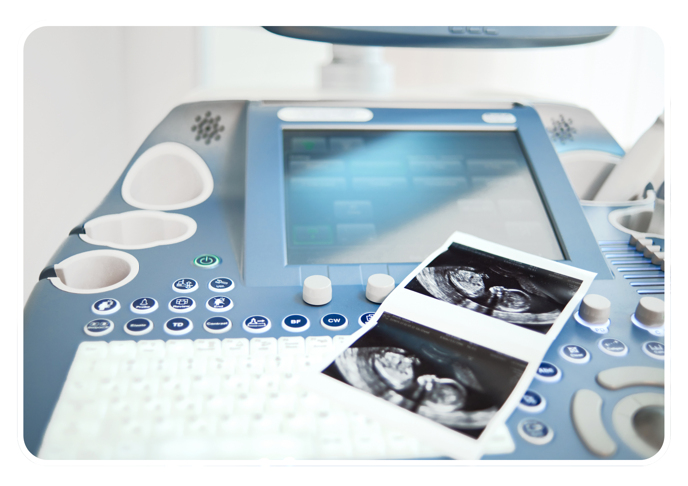
Sonography, or ultrasound, is a non-invasive method for monitoring fetal development. We provide high-quality ultrasound services.
Book Appointment

Sonography is a non-invasive and painless procedure that is often used to diagnose and monitor a variety of gynecological conditions, such as fibroids, ovarian cysts, endometriosis, and uterine abnormalities. It can also be used to monitor pregnancies and assess fetal development. It uses high-frequency sound waves to create images of the uterus, ovaries, and other pelvic organs, allowing physicians to visualize and evaluate the structure and function of these organs. Sonography is an important diagnostic tool that may evaluate an assortment of organs and systems, including the heart, arteries, female genitalia, belly, breasts, prostate, and other structures that may be suggested by clinical signs. At our sonography center, we understand that undergoing medical imaging tests can be stressful and intimidating. That’s why we prioritize patient comfort and care throughout the entire imaging process. Our team is dedicated to ensuring that patients feel comfortable and informed throughout the entire process, from check-in to receiving their results.
Gynecological 3D transvaginal sonography (TVS) is a high-resolution imaging technique used to visualize female reproductive organs, including the uterus and ovaries, for evaluating gynecological conditions. It provides detailed, volumetric images, but requires expertise in ultrasound and pelvic anatomy.
Transabdominal sonography, also known as abdominal ultrasound, is a non-invasive imaging technique that uses high-frequency sound waves to visualize internal organs and structures in the abdomen. It has clinical applications, including assessing organs, biliary systems, kidneys, and pelvic organs.
Obstetric 3D-4D sonography is a specialized imaging technique used during pregnancy to visualize and assess the developing fetus, providing detailed images for various clinical applications.
Fetal sonography, also known as fetal ultrasound, is a non-invasive imaging technique used during pregnancy to visualize and assess the developing fetus in utero. It provides valuable information about fetal growth, development, anatomy, and well-being. Frequency and timing depend on factors like maternal age and medical history.
An anomaly scan is a prenatal ultrasound examination performed between 18-22 weeks of pregnancy to detect potential fetal abnormalities, although it may not detect all abnormalities.
Color Doppler sonography is an advanced imaging technique used in ultrasound examinations to assess blood flow within organs, tissues, and vessels. It is useful for evaluating vascular structures and detecting abnormalities. It has clinical applications in vascular assessment, cardiac evaluation, obstetrics, and soft tissue imaging. However, it has limitations.