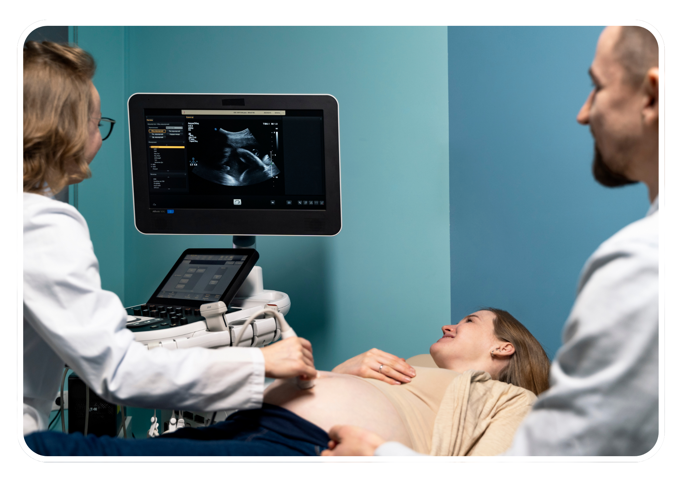
Prenatal Screening and Diagnosis
Obstetric sonography plays a crucial role in the prenatal screening and diagnosis of fetal abnormalities, genetic conditions, and pregnancy complications. Expert sonographers are trained to perform various types of ultrasound scans to assess fetal anatomy, growth, and well-being, and to detect any abnormalities or anomalies that may require further evaluation or intervention.
Routine Obstetric Ultrasound Scans
Dating Scan This ultrasound scan is performed early in pregnancy to accurately determine the gestational age of the fetus and estimate the due date. It helps establish the timing of prenatal care and screening tests.
Nuchal Translucency (NT) Scan The NT scan is a specialized ultrasound scan performed around 11-14 weeks of pregnancy to assess the thickness of the fluid at the back of the fetal neck. An increased NT measurement may indicate an increased risk of chromosomal abnormalities, such as Down syndrome.
Anomaly Scan (Detailed Fetal Anomaly Ultrasound) The anomaly scan, typically performed around 18-20 weeks of pregnancy, is a comprehensive ultrasound examination that evaluates fetal anatomy and development. It assesses the structure and function of various fetal organs and systems, including the brain, heart, spine, limbs, and abdominal organs, to detect any abnormalities or congenital anomalies.
Growth Scan Growth scans are performed periodically throughout pregnancy to monitor fetal growth and development. They assess parameters such as fetal size, weight, head circumference, abdominal circumference, and femur length, and compare them to expected growth standards for gestational age.
Fetal Well-Being Assessment / Colour Doppler Scan In late pregnancy, ultrasound scans may be used to assess fetal well-being by evaluating parameters such as amniotic fluid volume, fetal movements, and fetal heart rate patterns. These assessments help identify any signs of fetal distress or complications that may require further monitoring or intervention.
Specialized Obstetric Ultrasound
Fetal Echocardiography Fetal echocardiography is a specialized ultrasound examination of the fetal heart to assess cardiac anatomy and function. It is performed to diagnose congenital heart defects and other cardiac abnormalities.
3D/4D Ultrasound Three-dimensional (3D) and four-dimensional (4D) ultrasound imaging techniques provide detailed, three-dimensional images of the fetus, allowing for better visualization of fetal anatomy and features. 4D ultrasound adds the element of real-time motion, enabling parents to see dynamic images of their baby in utero.
Fetal Doppler Ultrasound Fetal Doppler ultrasound is used to assess blood flow in the umbilical cord, placenta, and fetal vessels. It helps evaluate fetal well-being and detect signs of fetal distress or intrauterine growth restriction (IUGR).
Guidance for Invasive Procedures Expert obstetric sonographers may also assist obstetricians and maternal-fetal medicine specialists during invasive procedures such as chorionic villus sampling (CVS), amniocentesis, fetal blood sampling, and fetal therapy interventions. Ultrasound guidance ensures accurate placement of the needle or instrument and reduces the risk of complications.
Importance and Benefits
Early Detection and Diagnosis Expert obstetric sonography is crucial for the early detection and diagnosis of fetal abnormalities and complications. This allows for timely intervention and management, improving pregnancy outcomes.
Enhanced Prenatal Care By providing detailed and accurate information about fetal development and maternal health, expert sonography supports comprehensive prenatal care, enabling healthcare providers to tailor their approach to each individual pregnancy.
Parental Reassurance and Bonding Seeing detailed images of their developing baby helps reassure expectant parents about the health and well-being of their child. Real-time 4D imaging enhances the emotional connection between parents and their unborn baby.
Support for High-Risk Pregnancies Expert sonography is especially important in managing high-risk pregnancies, providing critical information that guides the care and monitoring of pregnancies with potential complications.
Overall, expert obstetric sonography is a vital component of prenatal care, offering invaluable insights into fetal health and development and supporting informed decision-making and management of pregnancy. Collaboration between obstetric sonographers, obstetricians, maternal-fetal medicine specialists, and other healthcare professionals ensures comprehensive and effective prenatal care for pregnant individuals and their babies.

