Clinical Applications
Fetal sonography has various clinical applications in prenatal care, including:
- Dating and Viability: It is used to confirm pregnancy viability, determine gestational age, and estimate the due date (estimated date of delivery, EDD) based on fetal measurements such as crown-rump length (CRL) or biparietal diameter (BPD).
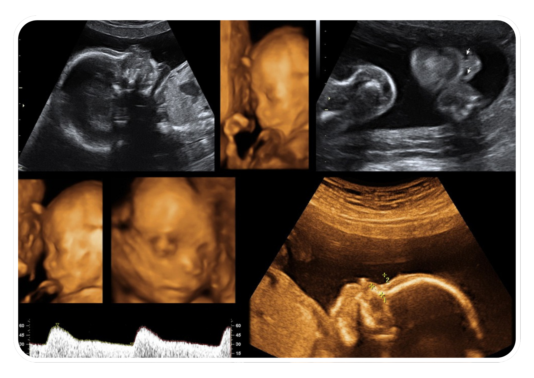
- Fetal Anatomy Scan: Fetal sonography is used to perform detailed fetal anatomy scans (anomaly scan) to assess the structure and development of the fetus. It allows for the visualization of fetal organs, limbs, spine, brain, heart, and other anatomical features to detect any abnormalities or congenital anomalies.
- Fetal Growth Assessment: It can monitor fetal growth and development throughout pregnancy by measuring fetal biometric parameters such as head circumference, abdominal circumference, femur length, and estimated fetal weight (EFW). Serial fetal sonography scans are performed to track fetal growth patterns and assess for fetal growth restriction or macrosomia.
- Amniotic Fluid Assessment: Fetal sonography is used to evaluate the quantity and quality of amniotic fluid surrounding the fetus. Abnormalities in amniotic fluid volume, such as oligohydramnios (decreased fluid) or polyhydramnios (increased fluid), may indicate underlying fetal or maternal conditions.
- Placental Assessment: It can assess the location, size, thickness, and vascularity of the placenta and detect placental abnormalities such as placenta previa (low-lying placenta) or placental insufficiency.
- Fetal Well-Being Assessment: Fetal sonography is used to assess fetal well-being by evaluating parameters such as fetal movements, fetal heart rate patterns (cardiotocography), and umbilical artery Doppler velocimetry. These assessments help determine fetal health, vitality, and response to intrauterine conditions.
Timing and Frequency
Fetal sonography is typically performed at different stages of pregnancy:
- First Trimester: Dating and viability scan to confirm the pregnancy, estimate the due date, and assess early fetal development.
- Second Trimester: Anomaly scan to assess fetal anatomy and detect congenital anomalies.
- Third Trimester: Fetal growth and well-being assessment to monitor growth patterns, amniotic fluid levels, and fetal position.
The frequency and timing of fetal sonography scans may vary depending on factors such as maternal age, medical history, pregnancy complications, and clinical indications.
Interpretation and Reporting
The images obtained from fetal sonography are interpreted by trained healthcare providers, including radiologists, obstetricians, sonographers, or other specialists. They analyze the images for abnormalities, document findings in a structured report, and communicate results to referring healthcare providers for further evaluation and management.
Procedure and Safety
- Preparation: No special preparation is usually required, but a full bladder may be needed for early pregnancy scans to improve image clarity.
- Procedure: A gel is applied to the abdomen to improve sound wave transmission, and a handheld transducer is moved over the skin to capture images.
- Safety: Fetal sonography is considered safe for both the mother and fetus, with no known harmful effects when performed appropriately.
Benefits
- Non-Invasive: Provides critical information without the need for invasive procedures.
- Real-Time Imaging: Allows for real-time assessment of fetal movements, heart rate, and other vital parameters.
- Early Detection: Facilitates early detection and management of fetal abnormalities and pregnancy complications.
- Parental Bonding: Enhances parental bonding by providing visual images of the developing fetus.
Conclusion
Fetal sonography is a valuable tool in prenatal care that provides essential information about fetal health, development, and well-being. It plays a crucial role in the assessment and management of pregnancy complications, fetal anomalies, and maternal-fetal conditions, contributing to improved pregnancy outcomes and patient care. By offering detailed insights into fetal development and identifying potential issues early, fetal sonography helps healthcare providers ensure the best possible outcomes for both mother and baby.