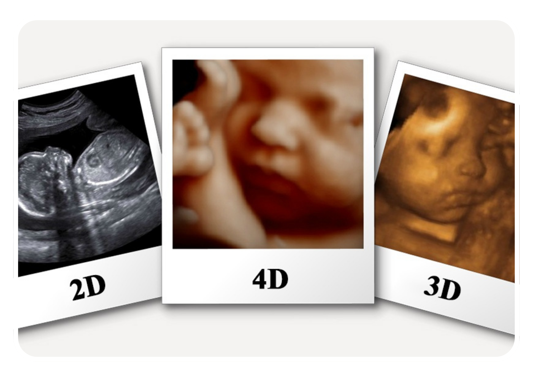
Technology and Imaging Process
Obstetric 3D-4D sonography utilizes advanced ultrasound technology to generate detailed, three-dimensional images of the fetus in utero. The process involves capturing multiple two-dimensional (2D) ultrasound images from different angles and orientations and then reconstructing them into three-dimensional images using specialized software. Real-time imaging (4D ultrasound) allows for the visualization of fetal movements and behaviors, providing dynamic video sequences of the fetus in motion.
Enhanced Visualization
Obstetric 3D-4D sonography offers enhanced visualization of fetal anatomy and features compared to traditional 2D ultrasound. It allows healthcare providers and expectant parents to see detailed images of the fetus's face, limbs, fingers, toes, and other structures with greater clarity and depth. Additionally, real-time 4D ultrasound provides dynamic views of fetal movements, facial expressions, and interactions in the womb, enhancing the prenatal bonding experience for parents.
Clinical Applications
Obstetric 3D-4D sonography has various clinical applications in prenatal care, including:
- Fetal Anatomy Scan: It is used to perform detailed fetal anatomy scans to assess the structure and development of the fetus. 3D-4D sonography allows for better visualization of fetal organs, limbs, facial features, and other anatomical details, aiding in the detection of fetal abnormalities or congenital anomalies.
- Fetal Growth Assessment: It can be used to monitor fetal growth and development throughout pregnancy by measuring parameters such as fetal size, weight, head circumference, abdominal circumference, and femur length. 3D-4D sonography provides accurate measurements and visualizations of fetal growth patterns.
- Fetal Well-Being Assessment: It is utilized to assess fetal well-being by evaluating parameters such as amniotic fluid volume, fetal movements, and fetal heart rate patterns. Real-time 4D ultrasound allows for the observation of fetal movements and behaviors, providing reassurance about fetal health and vitality.
- Prenatal Bonding: Obstetric 3D-4D sonography enhances the prenatal bonding experience for expectant parents by allowing them to see dynamic images and videos of their unborn baby in utero. Parents can observe fetal movements, facial expressions, and interactions, fostering a stronger emotional connection with their baby before birth.
Procedure and Safety
Obstetric 3D-4D sonography is typically performed by trained sonographers or healthcare providers in clinical settings, such as obstetric ultrasound departments or prenatal imaging centers. The procedure is safe and non-invasive, with no known risks to the pregnant individual or the fetus. A water-based gel is applied to the abdomen to improve acoustic coupling, and a handheld transducer is used to obtain images of the fetus. The examination is usually well-tolerated by pregnant individuals and does not require anesthesia or sedation.
Limitations
While obstetric 3D-4D sonography offers valuable information and insights into fetal development, it also has limitations. The quality of images may be affected by factors such as fetal position, maternal body habitus, amniotic fluid volume, and fetal movement. Additionally, 3D-4D sonography should be used judiciously and in conjunction with other prenatal screening and diagnostic tests to ensure comprehensive fetal assessment and accurate diagnosis.
Conclusion
Obstetric 3D-4D sonography is a valuable imaging modality in prenatal care that provides enhanced visualization of the fetus and enriches the prenatal bonding experience for expectant parents. When used appropriately by trained healthcare providers, it can contribute to improved fetal assessment, early detection of abnormalities, and parental engagement during pregnancy.